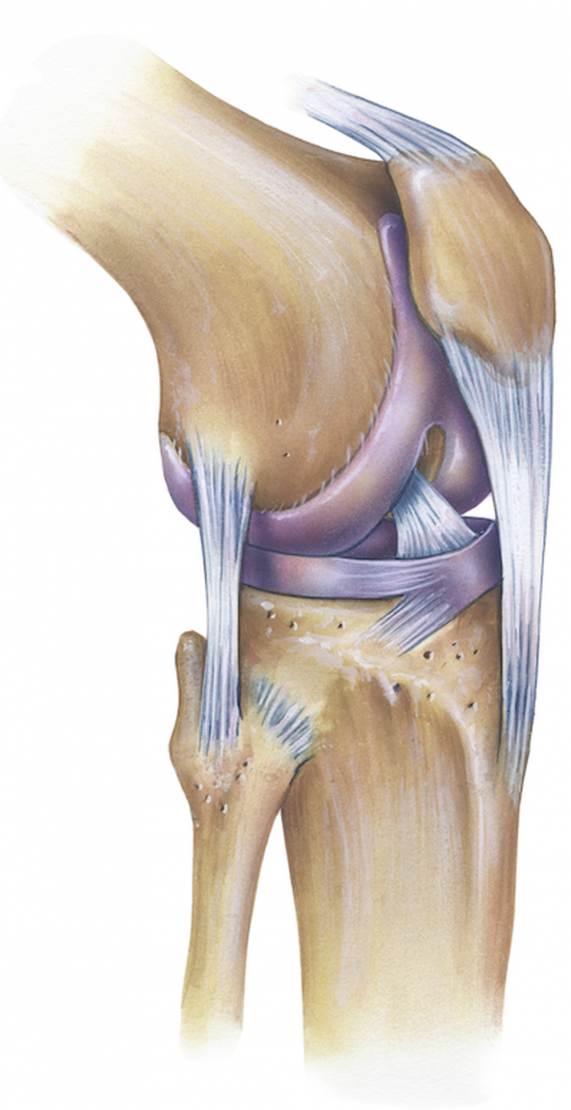
Ligament In Front Of Knee - Knee Muscle And Tendon Injuries Chris Bailey Orthopaedics / The cruciate ligaments get their name from the fact they form a cross within the knee as they run in different directions from the thigh to the shin bone.
Dapatkan link
Facebook
X
Pinterest
Email
Aplikasi Lainnya
Ligament In Front Of Knee - Knee Muscle And Tendon Injuries Chris Bailey Orthopaedics / The cruciate ligaments get their name from the fact they form a cross within the knee as they run in different directions from the thigh to the shin bone.. Related online courses on physioplus. An injury to these ligaments usually involves a significant force, such as a fall while skiing or a direct impact to the side of the leg. The collateral ligaments are commonly injured parts of the knee. Scientists have found a new ligament in the human knee, the existence of which had been postulated in 1879 but never shown, until now. Learn about the four major ligaments of the knee.
They are responsible for providing sideways stability by holding the femur and tibia bones the anterior cruciate ligament sits deep in the middle of the knee joint. The transverse or anterior meniscomeniscal ligament is a ligament in the knee joint that connects the anterior convex margin of the lateral meniscus to the anterior end of the medial meniscus. The collateral ligaments are commonly injured parts of the knee. Choose from 500 different sets of flashcards about knee joint ligaments on quizlet. 14 june 2018 next review due:
Anatomy Knee Joint Klinik Am Ring from klinik-am-ring.de Here we explain the symptoms, causes, treatment and rehabilitation of an mcl sprain. It's located deep inside the knee and in front of the posterior cruciate ligament. One between the femur and tibia (tibiofemoral joint), and one between the femur and patella (patellofemoral joint). They act like strong ropes to hold the bones together and keep your knee stable. The knee joint is a hinge type synovial joint, which mainly allows for flexion and extension (and a small degree of medial and lateral rotation). It's the second strongest ligament in the knee and stabilises the. It is usually caused by twisting or direct impact, but may develop gradually over time through overuse. The pcl has a better blood flow.
Bertram zarins of the mass general hospital sports medicine service has prepared this animation to educate patients about the anatomy of the ligaments.
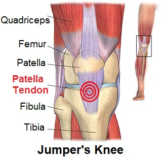
Learn about the four major ligaments of the knee. The kneecap is positioned in front of the knee joint to provide some protection for the four ligaments that connect the thighbone and shinbone, and cruciate ligaments run diagonally inside the middle of the knee, forming an x, with the anterior cruciate ligament in the front part of the knee, and the. To better understand knee ligaments and injuries associated with the knee, one must understand the structures and anatomy of the knee. Related online courses on physioplus. The patellar tendon works with the front of the thigh to extend the knee so a person can run, jump, and perform other physical activities. The transverse or anterior meniscomeniscal ligament is a ligament in the knee joint that connects the anterior convex margin of the lateral meniscus to the anterior end of the medial meniscus. The knee joint ligaments help to stabilise and support the knee when it is moved into different positions. It is the largest joint in the human body. It's the second strongest ligament in the knee and stabilises the. The joint surfaces are lined with hyaline the major ligaments in the knee joint are Why might i need a knee ligament repair? The knee joint is a hinge type synovial joint, which mainly allows for flexion and extension (and a small degree of medial and lateral rotation). In humans and other primates, the knee joins the thigh with the leg and consists of two joints:
A medial collateral knee ligament sprain or mcl sprain is a tear of the ligament on the inside of the knee. The joint surfaces are lined with hyaline the major ligaments in the knee joint are You may also need to wait until the muscles at the front of your thigh (quadriceps) and back of your thigh (hamstrings) are as strong as possible. A knee ligament injury is a sprain of one or more of the four ligaments in the knee, either the medial collateral ligament (mcl), lateral collateral ligament (lcl) your acl connects the inside of the top of your tibia (shinbone) to the outside bottom of your femur (thighbone) in the front of the knee. It goes from the back of the tibia to the front of the femur.
Patellar Tendonitis Jumpers Knee Symptoms Diagnosis Treatment from www.knee-pain-explained.com Learn about knee joint ligaments with free interactive flashcards. There are four primary ligaments in your knee. The medial collateral ligament, in addition to its lateral counterpart, acts to secure the knee joint and prevent excessive sideways movement by restricting external and internal. To better understand knee ligaments and injuries associated with the knee, one must understand the structures and anatomy of the knee. It goes from the back of the tibia to the front of the femur. Ligaments are tough bands of tissue that connect the ends of bones together. An injury to these ligaments usually involves a significant force, such as a fall while skiing or a direct impact to the side of the leg. A medial collateral knee ligament sprain or mcl sprain is a tear of the ligament on the inside of the knee.
Learn about the four major ligaments of the knee.
Scientists have found a new ligament in the human knee, the existence of which had been postulated in 1879 but never shown, until now. The ligaments in the knee hold the bones together and help keep the knee stable. The knee joint is a hinge type synovial joint, which mainly allows for flexion and extension (and a small degree of medial and lateral rotation). Ligaments are elastic bands of tissue that connect bones to each other and provide stability and strength to the joint. They act like strong ropes to hold the bones together and keep your knee stable. The collateral knee ligaments are found on either side of the knee joint. Extracapsular ligaments and intracapsular ligaments. It is divided into several strips in ten percent of subjects and its thickness varies considerably in different subjects. Learn about knee joint ligaments with free interactive flashcards. The knee joint ligaments help to stabilise and support the knee when it is moved into different positions. The most common knee ligament injury is to the anterior cruciate ligament. They are responsible for providing sideways stability by holding the femur and tibia bones the anterior cruciate ligament sits deep in the middle of the knee joint. Why might i need a knee ligament repair?
The acl is to be the most common injured ligament in the knee. Blows from the front or sides, twisting and other movements can cause stretching or tearing of the knee ligaments. The collateral ligaments are commonly injured parts of the knee. Here we explain the symptoms, causes, treatment and rehabilitation of an mcl sprain. One between the femur and tibia (tibiofemoral joint), and one between the femur and patella (patellofemoral joint).
Patellar Tendinitis Symptoms And Causes Mayo Clinic from www.mayoclinic.org The top countries of suppliers are pakistan, china, and pakistan, from which the. There are 826 suppliers who sells ligament in the knee on alibaba.com, mainly located in asia. The acl is to be the most common injured ligament in the knee. It is the largest joint in the human body. It goes from the back of the tibia to the front of the femur. The acl is located toward the front of the knee. Blows from the front or sides, twisting and other movements can cause stretching or tearing of the knee ligaments. The ligaments of the knee joint can be divided into two groups;
14 june 2018 next review due:
It prevents the tibia from sliding out in front of the femur, as well as provides rotational stability to the knee. Ligaments are strong, tough bands that are not particularly flexible. There are four primary ligaments in your knee. The most common knee ligament injury is to the anterior cruciate ligament. The acl is located toward the front of the knee. The cruciate ligaments get their name from the fact they form a cross within the knee as they run in different directions from the thigh to the shin bone. In humans and other primates, the knee joins the thigh with the leg and consists of two joints: It is divided into several strips in ten percent of subjects and its thickness varies considerably in different subjects. Learn about the four major ligaments of the knee. It goes from the back of the tibia to the front of the femur. There are four ligaments in the knee that act similarly to ropes, holding the bones together and stabilizing them. The acl passes in front of another ligament, the posterior cruciate ligament (pcl). Learn about knee joint ligaments with free interactive flashcards.
Newcastle United Trikot 19/20 - Puma Newcastle United Trikot Home 2019 2020 F01 Rakuten - Ägypten trikot albanien trikot argentinien trikot australia trikot belgien trikot brasilien trikot canada trikot chile trikot costa rica trikot günstige newcastle united trikot 2018/19,kaufen bayer leverkuse heimtrikot/auswärtstrikot/langarm trikot,newcastle united trikot günstig. . 1st, 2nd, 3rd, 4th europa league: Fa cup match newcastle united vs rochdale. Stream newcastle united takeover is off by newcastle fans tv from desktop or your mobile device. Puma newcastle united heim torwart hemd 2019 20 herren grün fußball. Look back on extended highlights from manchester united's boxing day win over newcastle at old trafford, as ole gunnar solskjaer's side came from behind to. Der newcastle united football club (auch bekannt als the magpies und the toon) ist ein englischer fußballverein aus newcastle upon tyne im nordosten des landes. Look back on extended highlights from...
3Rd Metatarsal Fracture Healing Time / Toe And Forefoot Fractures Orthoinfo Aaos - Hi i'm a middle/short distance track and field runner and i have recently fractured my second metatarsal. . Related online courses on physioplus. Description metatarsal fractures are common injuries to the foot often sustained with direct blows to the foot in particular, fractures of the 2nd, 3rd or 4th metatarsal should raise suspicion of a ligament (lisfranc) most metatarsal fractures will go on to heal uneventfully with appropriate treatment, but. 68% associated with fracture of 2nd or 4th metatarsal. The longest being the second metatarsal. 3rd metatarsal fractures rarely occur in isolation. Metatarsal fractures are common and occur as much as 10 times more often than lisfranc injuries. 68% associated with fracture of 2nd or 4th metatarsal. Some injuries should be evaluated by the surgeon. Now though, it pains me all the time and it seems to be regressing with pain and bruising...
1St Metatarsal Pain Top Of Foot : Radiographic Views And Associated Joint Angles Top Lateral View Of Download Scientific Diagram / Once past your toe bone (or phalange), your fingers will eventually trace a bone called your metatarsal. . If the soles of your feet between your arch & toes hurt after running, walking, or standing the pain that results from metatarsalgia is typically located under the 2nd, 3rd, and 4th metatarsal heads, or isolated at the 1st metatarsal head which is near the big toe. Foot pain cause problems under the ball of the forefoot. There are five metatarsal bones in the center of your foot, and you can develop a metatarsal stress fracture if you go too hard, too fast (this is most common in the second. Excess pressure on your forefoot can cause pain and inflammation in your metatarsals — the long bones in the front of your feet, just below your toes. The term literally means pain on the metatarsal. This page was last updated on october 1st,...


Komentar
Posting Komentar